
Blog · 1 July 2024 · 5 min read
Becoming a Slit Lamp Expert
1. Introduction to Slit Lamp Examination
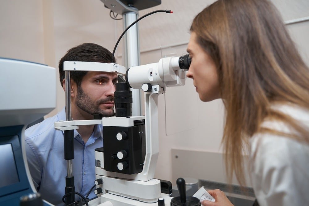
In the realm of eye care, the slit lamp examination stands as a fundamental diagnostic tool, offering detailed insights into various ocular conditions. This article explores the components, procedure, diagnostic capabilities, advantages, limitations, and future trends associated with slit lamp examinations.
2. Components of a Slit Lamp
Illumination System
The slit lamp’s illumination system plays a crucial role in providing a concentrated and adjustable light beam. This illumination is essential for illuminating specific areas of the eye during examination, aiding in the detection of abnormalities such as corneal abrasions or foreign bodies.
Binocular Microscope
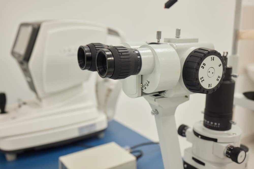
Integrated with the illumination system, the binocular microscope of the slit lamp provides stereoscopic views of the eye’s structures. This dual-view system allows clinicians to observe the depth and dimension of ocular tissues with enhanced clarity and depth perception.
Chin Rest and Headrest
Patients rest their chin and forehead on a stable chin rest and headrest during the examination. This positioning ensures the patient’s comfort and maintains proper alignment relative to the slit lamp, facilitating consistent and accurate examination results.
Joystick and Fine Adjustment Controls
Control mechanisms like the joystick and fine adjustment knobs enable precise manipulation of the slit lamp’s light beam and microscope. Clinicians use these controls to adjust the focus, angle, and intensity of the illumination, optimizing visualization of specific eye structures.
Procedure of a Slit Lamp Examination
Preparation of the Patient
Before starting the examination, clinicians explain the procedure to the patient, addressing any concerns and ensuring cooperation. Patients may need to remove contact lenses or eyeglasses and refrain from rubbing their eyes to facilitate clear examination.
Positioning and Alignment
Proper positioning of the patient relative to the slit lamp is critical for obtaining accurate and consistent results. Clinicians adjust the height and angle of the microscope and ensure the patient’s eyes are aligned with the light beam for optimal visualization.
Step-by-Step Examination Process
The slit lamp examination follows a systematic approach, examining various structures of the eye in detail:
Examination of the Eyelids and Adnexa
Clinicians assess the eyelids, eyelashes, and surrounding tissues for signs of inflammation, infection, or abnormalities.
Assessment of the Conjunctiva and Sclera
The slit lamp allows for detailed examination of the conjunctiva (the thin, transparent membrane covering the sclera) and sclera (the white outer layer of the eye), identifying conditions like conjunctivitis or scleritis.
Examination of the Cornea
Detailed inspection of the cornea involves observing its clarity, shape, and the presence of any irregularities such as scars, abrasions, or ulcers.
Inspection of the Anterior Chamber
The slit lamp enables clinicians to evaluate the anterior chamber of the eye, assessing the depth, clarity, and presence of cells or flares indicative of conditions like uveitis or glaucoma.
Evaluation of the Iris and Pupil
Examination of the iris includes assessing its color, shape, and pupillary reactions to light stimuli, providing insights into conditions like iritis or pupil abnormalities.
Examination of the Lens and Vitreous
The slit lamp facilitates detailed examination of the lens for opacity (cataracts) and visualization of the vitreous humor, aiding in the diagnosis of conditions affecting these structures.
Common Conditions Diagnosed with Slit Lamp Examination
Slit lamp examinations are instrumental in diagnosing various ocular conditions, including:
- Corneal Abrasions and Ulcers: Visible under slit lamp as disruptions in the corneal epithelium.
- Cataracts: Identified by opacities in the lens observed during examination.
- Conjunctivitis: Characterized by redness and inflammation of the conjunctiva.
- Glaucoma: Evaluated through assessment of the anterior chamber and optic nerve head.
- Diabetic Retinopathy: Signs such as microaneurysms or retinal hemorrhages are detectable.
Advantages of Slit Lamp Examination
The slit lamp offers several advantages in ocular diagnostics:
Detailed Visualization
By magnifying ocular structures, the slit lamp enables clinicians to detect subtle changes indicative of pathology, facilitating early diagnosis and intervention.
Precision Diagnosis
Clear and magnified views provided by the slit lamp aid in precise identification and characterization of ocular conditions, guiding appropriate treatment strategies.
Monitoring Progression
Regular slit lamp examinations allow for longitudinal assessment of eye conditions, monitoring progression, and evaluating treatment effectiveness over time.
Limitations and Considerations
Despite its benefits, slit lamp examinations have considerations:
Patient Cooperation
Patients need to remain still and cooperate during the examination to ensure accurate results, which can be challenging for some individuals, especially children or those with cognitive impairments.
Technical Expertise Required
Interpreting slit lamp findings requires specialized training and experience to distinguish between normal variations and pathological changes accurately.
Potential Discomfort for Patients
The proximity of the light source and microscope during slit lamp examinations may cause discomfort or sensitivity for some patients, although the procedure is generally well-tolerated.
Importance in Different Specialties
Slit lamp examinations are integral across various medical disciplines:
- Ophthalmology: Primary tool for diagnosing and managing eye diseases.
- Optometry: Essential for assessing refractive errors and detecting ocular abnormalities.
- Emergency Medicine: Facilitates rapid evaluation of eye injuries and acute conditions, guiding immediate treatment decisions.
Training and Certification
Proficiency in conducting slit lamp examinations requires formal education and ongoing training:
Educational Requirements
Ophthalmologists and optometrists undergo comprehensive training in slit lamp technology during their professional education, encompassing both theoretical knowledge and practical application.
Continuing Education for Practitioners
Continuous professional development ensures clinicians stay updated on advancements in slit lamp technology, diagnostic methodologies, and treatment modalities to enhance patient care.
Future Trends in Slit Lamp Technology
Advancements in slit lamp technology continue to shape the future of eye care:
Advancements in Imaging and Digital Integration
Integration of high-definition imaging systems enhances diagnostic capabilities, allowing for more detailed visualization of ocular structures and abnormalities.
Potential for Telemedicine Applications
The incorporation of slit lamp examinations into telemedicine platforms enables remote consultations, expanding access to specialized eye care services and facilitating timely interventions for patients worldwide.
Conclusion
The slit lamp examination remains indispensable in the field of eye care, offering unparalleled insights into ocular health. Its role in precision diagnostics and therapeutic monitoring continues to evolve, driving advancements in clinical practice and enhancing visual outcomes for patients globally.
Related Questions
What does a slit lamp examination involve?
A slit lamp examination involves using a specialized microscope to examine the structures of the eye in detail under magnification.
Is slit lamp examination painful?
No, slit lamp examinations are generally painless, although patients may experience slight discomfort due to the proximity of the instrument.
How long does a slit lamp examination take?
The duration of a slit lamp examination varies but typically lasts between 10 to 20 minutes, depending on the complexity of the evaluation required.
Can slit lamp examinations detect all eye conditions?
While slit lamp examinations are highly effective, some conditions may require additional imaging or specialized testing for comprehensive diagnosis.
More guides
All articles
7 September 2024 · 12 min read
The Future of Eye Care: Tele-Optometry and Remote Care
This comprehensive article explores the rise of tele-optometry, The article also delves into the technology behind tele-optometry,

30 August 2024 · 12 min read
The Importance of Objective Refraction in Optometry
What objective refraction is, how autorefractors and retinoscopes measure refractive error without patient feedback, and when it matters most.
9 August 2024 · 7 min read
Guide to Understanding the Benefits of an Automatic Keratometer
How an automatic keratometer measures corneal curvature, where it helps (astigmatism, keratoconus, contact-lens fitting) and how to choose one.